
![]()
![]()
![]()
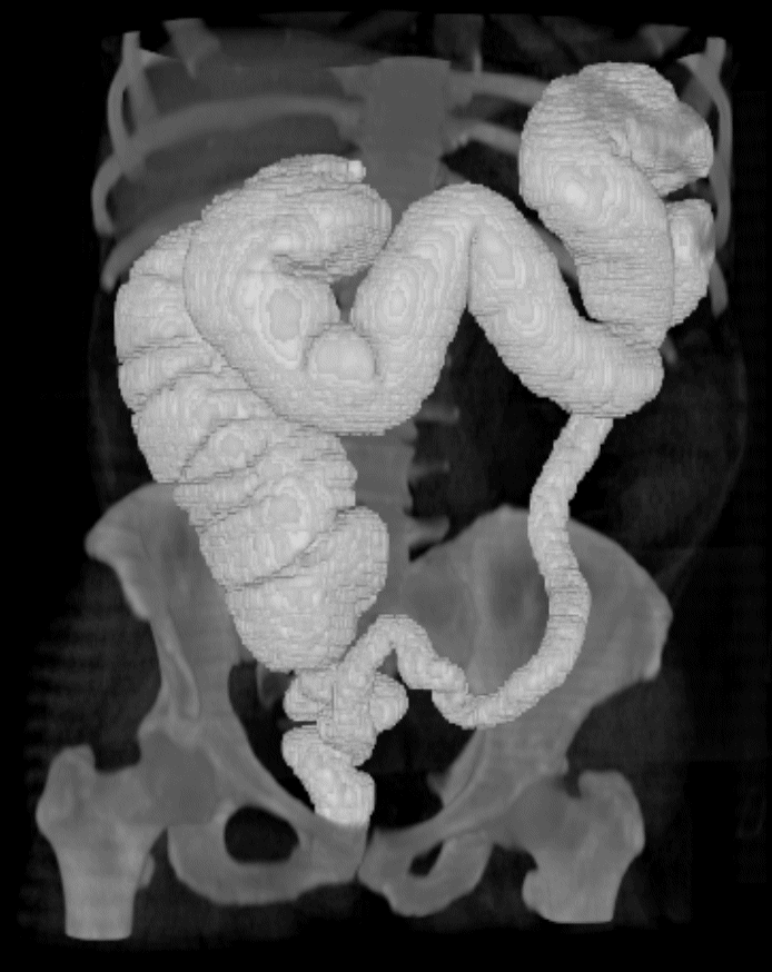 3D image of the Colon - Click on Image to see the movie
|
This is an new emerging
technique devised to replace the conventional Colonoscopy.
It uses a computed tomography (CT) scanner. This CT scanner uses X-rays to recreate a 3D model of the colon. This technique is non-invasive. When it is applied to the patients abdomen it generates the data necessary to create a virtual reality model of the colon. This scanning is done while the patient is lying face down and when they are lying face up. Scanning is done during a single breath-hold to reduce any movement. The scanner takes many image slices of the colon. These slices are virtual axial cuts of the body displaying internal density values. These slices can be combined together to generate a 3D density map of a scanned volume Since the preparation for this examination is the same as for the Colonoscopy it is well suited for screening an air filled colon as there is a high contrast between air and soft tissue densities. The contrast between the tissue and the cavity of the colon
allow the radiologist to distinguish between them, therefore increasing
the chances of detecting any abnormalities.
Note: Density is
measured in Hounsfield Units (HU) after Hounsfield as he introduced the
technique by which X-ray transmission readings are taken through the
object being examined at a multitude of angles
|